

For most pregnant patients, prenatal care is focused appropriately on the pregnancy: fetal development, maternal physiology, hypertension, diabetes, infection, genetic abnormalities, and obstetric complications.
Cancer is rarely near the top of the list.
That creates a paradox.
Pregnancy brings a woman into repeated contact with the healthcare system. She may have a dozen or more clinical encounters. She undergoes physical examinations, blood testing, cervical assessment, ultrasound imaging, and, increasingly, cell-free DNA testing. Yet malignancy can still be missed because symptoms are attributed to pregnancy, clinicians hesitate to order imaging, and laboratory abnormalities are assumed to be physiologic.
The result is that pregnancy can simultaneously create opportunities for earlier cancer detection and conditions for delayed diagnosis.
And a remarkable new development has added another dimension: a blood test ordered to examine fetal chromosomes can occasionally detect genomic evidence of an unsuspected maternal cancer.
The implications go far beyond obstetrics.
How common is cancer during pregnancy?
Cancer complicating pregnancy remains uncommon, but it is not extraordinarily rare.
Population-based studies generally place pregnancy-associated cancer in the neighborhood of 1 per 1,000 pregnancies, although estimates vary substantially depending on whether investigators count cancers diagnosed only during pregnancy or extend the definition to 6, 12, or even 24 months postpartum. A systematic review found that only about one quarter of pregnancy-associated cancers were actually diagnosed during pregnancy, with many identified after delivery. [1,2]
In a U.S. population-based study of 775,709 pregnancies, the incidence of cancer diagnosed during pregnancy or within one year postpartum was approximately 109 per 100,000 pregnancies. The most frequent invasive cancers were:
breast cancer
thyroid cancer
melanoma
hematologic malignancies
cervical and uterine cancers. [2]
Different populations produce somewhat different rankings. Swedish data found melanoma, breast cancer, and cervical cancer to be the three most common malignancies diagnosed during pregnancy. More recent Swedish data likewise showed melanoma, breast, and cervical cancer accounting for more than half of cancers detected during pregnancy. [3,4]
One reason the issue is becoming more important is straightforward: maternal age at pregnancy is increasing, while the incidence of several cancers, including breast and colorectal cancer, rises with age.
The first danger: symptoms of cancer can look like pregnancy
Cancer does not become biologically invisible during pregnancy. It becomes clinically easier to explain away.
Consider some examples:
A breast lump may be attributed to normal gestational breast change.
Fatigue may be attributed to pregnancy.
Anemia may be attributed to iron deficiency.
Rectal bleeding may be attributed to hemorrhoids.
Constipation may be attributed to progesterone.
Abdominal distention may be attributed to the enlarging uterus.
Nausea and weight change may be attributed to pregnancy.
Dyspnea may be attributed to normal cardiopulmonary adaptation.
Headache may be attributed to migraine or hypertensive disease.
Lymphadenopathy may initially be observed rather than biopsied.
Abnormal vaginal bleeding may be attributed to pregnancy itself.
This phenomenon is particularly concerning with colorectal cancer. Pregnancy-associated colorectal cancer is often diagnosed relatively late, in part because rectal bleeding, constipation, abdominal pain, anemia, and altered bowel habits have plausible obstetric explanations. [5,6]
The clinical rule therefore should not be:
“Could pregnancy explain this symptom?”
Of course it could.
The better question is:
“Is the severity, persistence, progression, or pattern of this symptom adequately explained by normal pregnancy?”
That is a very different threshold.
Pregnancy does not prohibit appropriate cancer imaging
There remains considerable confusion among both patients and clinicians regarding imaging during pregnancy.
Ultrasound and MRI without contrast are generally preferred first-line techniques because they do not use ionizing radiation. But that does not mean that CT, radiography, nuclear medicine, contrast-enhanced imaging, or PET are universally forbidden.
The 2026 Society for Maternal-Fetal Medicine Consult Series on cancer in pregnancy recommends ultrasound and noncontrast MRI as first-line modalities. Importantly, however, it states that CT with or without contrast, gadolinium-enhanced MRI, and FDG-PET/CT should not be withheld when they are clinically indicated. [7]
ACOG has made the same broader principle clear: most diagnostic radiographic procedures expose the fetus to doses far below thresholds associated with fetal harm, and necessary imaging should not be withheld simply because a patient is pregnant. Gadolinium should be restricted to situations in which it materially improves diagnosis and is expected to improve maternal or fetal outcome. [8]
That distinction matters enormously.
The relevant comparison is not:
radiation versus no radiation.
It is:
the potential fetal risk of the diagnostic procedure versus the maternal and fetal risk of failing to diagnose a potentially lethal disease.
Which cancers can actually be detected during pregnancy?
Almost any cancer can be diagnosed during pregnancy.
That does not mean pregnancy provides validated screening for most cancers.
The important distinction is between:
routine screening that remains appropriate during pregnancy
incidental detection during obstetric care
diagnostic evaluation prompted by symptoms or abnormal findings
emerging genomic signals, especially abnormal prenatal cfDNA
The practical landscape looks like this:
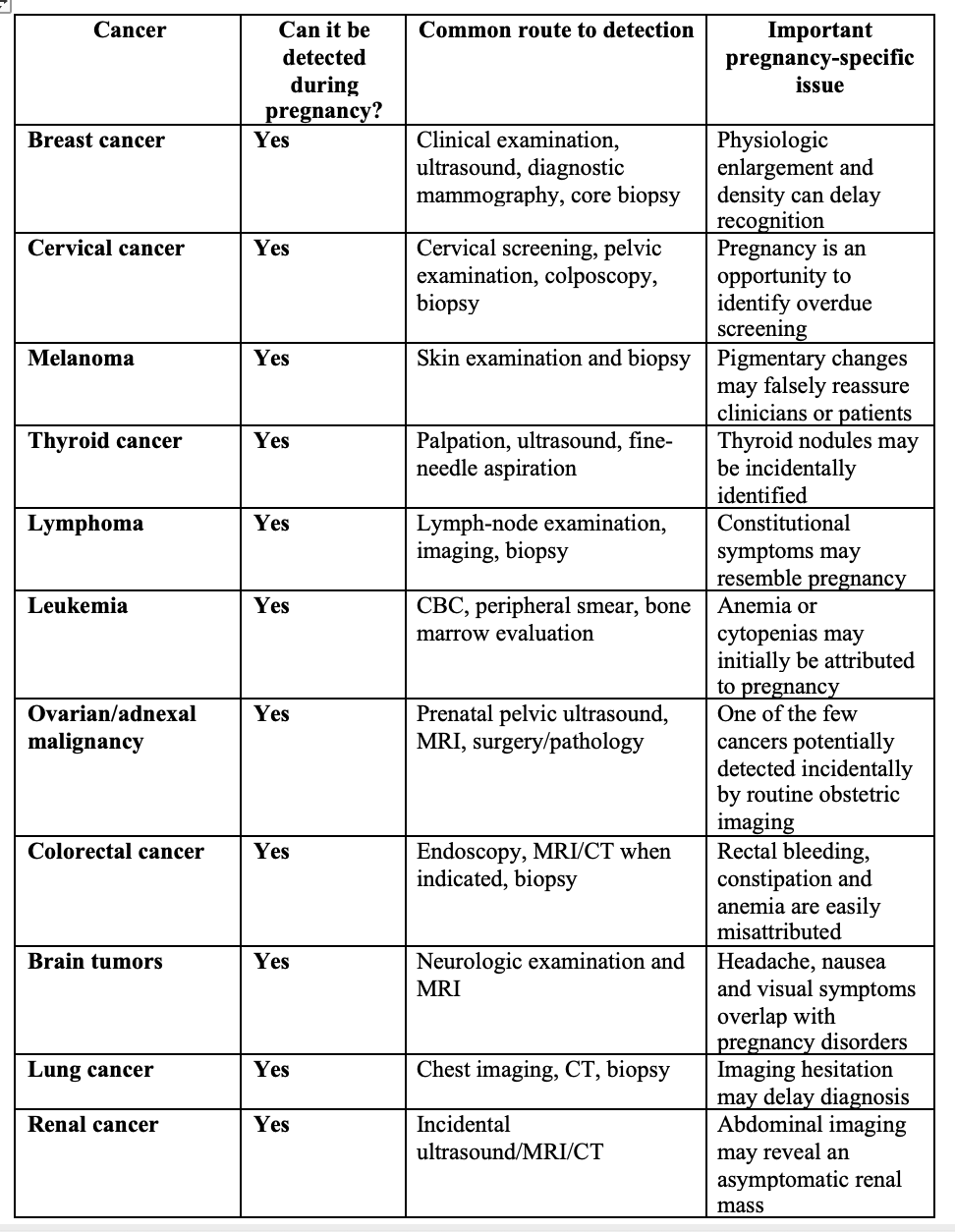
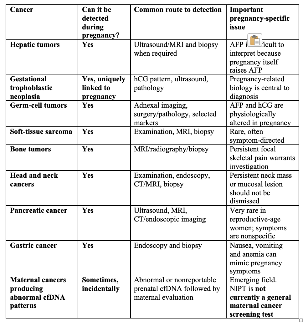
This table makes an important point:
The question is usually not whether a cancer can be diagnosed during pregnancy.
It can.
The larger question is whether clinicians recognize the signal and pursue the diagnosis.
Breast cancer deserves special attention
Breast cancer is among the most frequent malignancies associated with pregnancy.
Pregnancy presents several diagnostic challenges. Breast volume increases, glandular tissue proliferates, breast density increases, and nodularity is common. These entirely normal physiologic changes make palpation and imaging interpretation more difficult.
But a persistent breast mass during pregnancy should not be observed indefinitely.
The American College of Radiology considers ultrasound usually appropriate as the initial study for a pregnant woman younger than 30 years with a palpable mass or focal breast symptom. When ultrasound demonstrates a suspicious lesion, diagnostic mammography and image-guided core biopsy can also be appropriate. [9]
Pregnancy is therefore not a reason to defer tissue diagnosis.
The central patient-safety issue is diagnostic delay.
Cervical cancer: prenatal care may provide a genuine screening opportunity
Unlike most malignancies, cervical cancer already has an established population screening infrastructure.
Pregnancy may bring previously underscreened women into healthcare and therefore provides an opportunity to determine whether cervical screening is due.
The 2026 ACOG recommendations endorse cervical cytology every three years for average-risk individuals aged 21 to 29, and primary high-risk HPV testing every five years for most individuals aged 30 to 65, with defined alternatives. [10]
Pregnancy does not erase the need to recognize a visibly abnormal cervix, unexplained bleeding, or abnormal screening result.
Here again, however, the purpose should not be to invent a special “pregnancy cancer screen.”
It should be to ensure that appropriate routine preventive care does not disappear simply because the patient is pregnant.
Ovarian/Fallopian tube cancer: prenatal ultrasound creates a special opportunity
Pregnancy is one of the few times when large numbers of otherwise healthy women undergo repeated pelvic imaging.
As a result, adnexal masses may be detected incidentally.
The overwhelming majority are benign, and ovarian/fallopian tube cancer during pregnancy is rare. But suspicious morphology requires appropriate evaluation.
Ultrasound is the primary modality for characterizing adnexal masses, with MRI useful when ultrasonography cannot adequately determine the nature or extent of a lesion. ACOG guidance on adnexal masses explicitly includes pregnant patients and emphasizes identifying lesions that warrant gynecologic-oncology consultation. [11]
This is genuine opportunistic detection.
It is very different from population ovarian-cancer screening.
Tumor markers become particularly treacherous in pregnancy
Pregnancy changes the interpretation of several markers commonly associated with malignancy.
AFP and hCG are obvious examples because they are integral components of pregnancy physiology.
CA-125 can also fluctuate, particularly early in pregnancy. Other markers including squamous-cell carcinoma antigen and CA 15-3 may show pregnancy-associated variation. [12,13]
Therefore:
An abnormal tumor marker during pregnancy is not equivalent to an abnormal tumor marker outside pregnancy.
Markers should generally support a clinical diagnosis rather than substitute for imaging and tissue pathology.
Then came NIPT: fetal testing unexpectedly begins detecting maternal cancer
This may be the most conceptually important development.
Noninvasive prenatal testing analyzes circulating cell-free DNA in maternal plasma.
Despite the familiar term cell-free fetal DNA, most DNA fragments circulating in maternal blood are actually maternal. The pregnancy-associated component comes predominantly from placental trophoblasts.
That means an abnormal maternal clone or cancer can sometimes alter the sequencing pattern.
And that is precisely what investigators began observing.
Some prenatal cfDNA tests produced bizarre results:
multiple aneuploidies,
multiple chromosomal gains and losses,
or results that could not plausibly be reconciled with an apparently normal fetus.
The explanation in a subset of patients was not fetal disease.
It was maternal malignancy.
The IDENTIFY study changed the discussion
In the NIH-led IDENTIFY study published in the New England Journal of Medicine, investigators evaluated pregnant or postpartum patients who had unusual or nonreportable prenatal cfDNA results that raised concern for maternal cancer.
The evaluation included rapid whole-body MRI, laboratory testing, and research genome-wide cfDNA sequencing.
Cancer was identified in 52 of 107 participants, 48.6%. [14]
Even more strikingly, 49 individuals had a genomic pattern involving multiple copy-number gains and losses across three or more chromosomes.
Cancer was found in 47 of those 49 patients, 95.9%. [14]
Whole-body MRI had a reported sensitivity of 98.0% and specificity of 88.5% within this highly selected cohort. Physical examination and conventional laboratory testing were considerably less informative. [14]
This is extraordinary.
But it must be interpreted correctly.
Forty-eight percent does NOT mean that 48% of women with an uninterpretable NIPT have cancer.
The study population was highly selected.
Patients with routine technical failures, such as low fetal fraction, were not equivalent to patients with highly abnormal genomic patterns suggestive of widespread chromosomal disruption.
A subsequent NEJM correspondence specifically emphasized this selection issue. [15]
So NIPT is not currently a validated maternal pan-cancer screening test.
But some highly abnormal cfDNA signatures clearly cannot safely be ignored.
The Society of Maternal-Fetal Medicine (SMFM) has now incorporated this into clinical guidance
The implication has already moved from research into clinical guidance.
The 2026 Society for Maternal-Fetal Medicine Consult Series on cancer in pregnancy recommends that maternal malignancy be considered when cfDNA screening demonstrates multiple chromosomal aneuploidies or a single autosomal monosomy that is discordant with fetal findings. [7]
That represents an important change in prenatal medicine.
A prenatal genetic test can now generate information relevant not only to:
the fetus
but potentially to:
the pregnant patient.
The ethical implications are substantial.
Pretest counseling traditionally focuses overwhelmingly on fetal genetic information.
But if genome-wide cfDNA platforms can generate clinically consequential maternal signals, patients need to understand that possibility.
Could prenatal cfDNA eventually become a maternal cancer screening test?
Possibly.
But we are not there yet.
There is enormous scientific interest in multi-cancer early detection using circulating tumor DNA. Pregnancy offers an unusual biological setting because millions of women already undergo cfDNA sequencing.
But several questions remain unresolved:
What genomic pattern should trigger evaluation?
What is the positive predictive value in an unselected obstetric population?
How often would evaluation generate false positives?
Which cancers are preferentially detected?
What should the diagnostic pathway be?
Should whole-body MRI become the standard confirmatory evaluation for a strongly suspicious maternal cfDNA pattern?
Who pays for that evaluation?
And perhaps most importantly:
Should a prenatal fetal screening test deliberately become a dual fetal-maternal genomic screening program?
That would require much stronger evidence than we currently possess.
Pregnancy may also change future cancer risk, but not in one direction
The relationship between pregnancy and cancer risk is often oversimplified.
Pregnancy does not uniformly “protect against cancer.”
Its effects vary substantially by cancer type and over time.
Breast cancer: first an increase, eventually a reduction
The relationship between childbirth and breast cancer is particularly interesting.
A large pooled analysis of 15 prospective studies, including more than 18,000 breast cancers, found that compared with nulliparous women, breast-cancer risk temporarily increased after childbirth, peaking roughly five years postpartum.
At that point the hazard ratio was approximately 1.80.
Risk then gradually declined, crossed below that of nulliparous women approximately 24 years after childbirth, and reached an HR of approximately 0.77 after 34 years. [16]
This is an important correction to the simplistic statement that pregnancy is immediately protective against breast cancer.
It is not.
The relationship is time dependent.
Pregnancy and postpartum breast involution produce profound remodeling of breast tissue. Those biological processes are under active investigation as possible contributors to the distinctive behavior of postpartum breast cancer.
Endometrial cancer: pregnancy is associated with substantially lower long-term risk
For endometrial cancer, the epidemiology is much more consistently protective.
A pooled analysis from the Epidemiology of Endometrial Cancer Consortium examined nearly 17,000 women with endometrial cancer and more than 39,000 controls.
Having had a full-term pregnancy was associated with an approximately 41% lower risk of endometrial cancer than never having had a full-term pregnancy, with an odds ratio of 0.59. Additional pregnancies were associated with further risk reduction. [17]
A separate meta-analysis similarly found substantially lower endometrial-cancer risk among parous compared with nulliparous women. [18]
These findings are observational and do not mean that pregnancy should ever be conceptualized as a cancer-prevention intervention.
But the association is strong and biologically plausible.
Ovarian cancer: parity is also associated with lower risk
Parity is one of the established epidemiologic factors associated with lower epithelial ovarian-cancer risk.
Large pooled analyses show progressively lower risk with greater parity, although the magnitude differs by histologic subtype.
For example, in the Ovarian Cancer Cohort Consortium, increasing parity was particularly strongly associated with lower risks of endometrioid and clear-cell ovarian cancers. [19]
The mechanism is probably not a single pathway. Reduced lifetime ovulation is likely part of the explanation, but hormonal and reproductive remodeling may contribute as well. [20]
Thyroid cancer: the evidence is weaker and less consistent
Several studies have examined whether pregnancy and parity influence thyroid-cancer risk.
A meta-analysis found only a modest association between ever having given birth and thyroid cancer, approximately RR 1.09, and importantly found no clear dose-response relationship. [21]
Another meta-analysis found no overall increase simply from a history of pregnancy, although some analyses suggested higher risk with multiple pregnancies or when thyroid cancer occurred within several years of pregnancy. [22]
These findings should therefore be interpreted cautiously.
They do not justify additional thyroid-cancer screening solely because a woman has been pregnant.
Melanoma: pregnancy itself has not been convincingly shown to worsen the disease
Melanoma is relatively common among cancers diagnosed in reproductive-age women and therefore appears frequently in pregnancy-associated cancer registries.
Historically, there was concern that pregnancy-related hormonal or immune changes might accelerate melanoma.
Modern data are considerably less convincing.
Recent reviews suggest that pregnancy itself does not reliably worsen melanoma-specific survival. When poorer outcomes are observed, delayed recognition, postpartum diagnosis, or more advanced stage at presentation may explain at least part of the difference. [23]
The lesson again is less about pregnancy causing aggressive cancer and more about diagnostic vigilance.
What pregnancy probably should NOT become
There is a danger in moving too far in the opposite direction.
Pregnancy should not become an excuse for indiscriminate whole-body cancer screening.
That would produce false positives, incidental findings, anxiety, procedures, cost, and potentially unnecessary intervention.
The evidence does not support routinely performing:
whole-body MRI,
pan-cancer tumor markers,
expanded CT screening,
or maternal cancer genomic screening
in every pregnant patient.
Most cancers in reproductive-age women remain uncommon.
A screening program requires evidence that testing improves meaningful outcomes and that benefits exceed harms.
That evidence does not yet exist for general maternal cancer screening during pregnancy.
But there is a large space between “screen everybody” and “ignore the possibility”
That middle ground is where clinical practice should improve.
We can do several things now.
First, persistent symptoms deserve investigation.
Pregnancy should modify the differential diagnosis.
It should not terminate it.
Second, necessary imaging should not be reflexively withheld.
Ultrasound and MRI are often excellent first-line tools, but CT and other modalities remain appropriate when the clinical benefit justifies them. [7,8]
Third, tissue diagnosis remains decisive.
Suspicious breast masses, lymph nodes, skin lesions, cervical lesions, or other accessible abnormalities should be biopsied when clinically indicated.
Fourth, use routine obstetric imaging intelligently.
An adnexal lesion, renal mass, hepatic lesion, or other unexpected finding deserves structured follow-up rather than dismissal as an incidental curiosity.
Fifth, recognize abnormal cfDNA patterns as potentially maternal.
A result suggesting multiple chromosomal abnormalities that cannot be explained by fetal testing is no longer merely an “NIPT failure.”
It may be a maternal cancer signal. [7,14]
We may need a new concept: maternal incidental genomic findings
Prenatal testing was built conceptually around the fetus.
That model is becoming obsolete.
Modern cfDNA sequencing simultaneously samples information derived from the placenta and the pregnant patient.
As sequencing becomes broader and more sensitive, it may reveal:
maternal chromosomal abnormalities,
clonal hematopoiesis,
maternal copy-number variants,
fibroids,
organ transplantation,
and occasionally cancer.
The prenatal genetic test is therefore becoming a maternal-fetal genomic test whether we intended it to or not.
That creates a responsibility to establish clear pathways for disclosure, counseling, confirmatory testing, oncology referral, and long-term follow-up.
And what about the placenta?
There is another clinically important point that receives surprisingly little attention.
SMFM now recommends placental pathology examination in all pregnancies complicated by maternal cancer, regardless of cancer type or treatment. [7]
Maternal malignancies rarely metastasize to the placenta or fetus, but placental examination can provide clinically important information when cancer occurs during pregnancy.
This is precisely the type of issue that can fall between oncology, obstetrics, pathology, neonatology, and pediatrics unless responsibility is explicitly assigned.
The larger patient-safety lesson
Cancer during pregnancy exposes a familiar weakness in medicine.
When a common explanation exists, clinicians may stop searching.
Pregnancy provides hundreds of common explanations.
Fatigue.
Anemia.
Constipation.
Breast changes.
Bleeding.
Nausea.
Weight change.
Pain.
Dyspnea.
Headache.
Each is usually benign.
Occasionally, it is not.
The goal cannot be to investigate every symptom as cancer.
But neither should pregnancy become a diagnostic shield behind which malignancy remains invisible.
The future may be very different
Within the next decade, the concept of prenatal testing may expand considerably.
A maternal blood sample obtained for fetal screening could theoretically provide information about:
fetal chromosome abnormalities,
maternal genetic disease,
placental dysfunction,
preeclampsia risk,
preterm-birth risk,
and potentially occult maternal cancer.
That future is scientifically plausible.
It is not yet clinically validated.
The distinction matters.
We should resist both extremes: dismissing the technology because it is immature, and deploying it clinically before we know whether it improves outcomes.
The strongest evidence today supports a narrower conclusion.
Pregnancy is not a general cancer-screening program.
But pregnancy provides an unusually intense window of contact with the healthcare system.
We should use that window intelligently.
Persistent or unusual symptoms should be investigated.
Age-appropriate cancer screening should not disappear.
Suspicious imaging findings require follow-up.
Necessary diagnostic procedures should not be withheld simply because the patient is pregnant.
And highly abnormal prenatal cfDNA findings that do not correspond to the fetus may represent something we once never imagined finding in a prenatal genetics laboratory:
the mother’s cancer.
That changes prenatal diagnosis.
And it may eventually change cancer detection as well.
References
de Haan J, Verheecke M, Van Calsteren K, et al. Oncological management and obstetric and neonatal outcomes for women diagnosed with cancer during pregnancy: a 20-year international cohort study. Lancet Oncol. 2018;19:337-346.
Cottreau CM, Dashevsky I, Andrade SE, et al. Pregnancy-associated cancer: a U.S. population-based study. J Womens Health. 2019;28:250-257.
Andersson TM, Johansson ALV, Fredriksson I, Lambe M. Cancer during pregnancy and the postpartum period: a population-based study. Cancer. 2015;121:2072-2077.
Lu D, Ludvigsson JF, Smedby KE, et al. Risk factors for the increasing incidence of pregnancy-associated cancer in Sweden: a population-based study. Acta Obstet Gynecol Scand. 2023.
Paspulati RM, et al. Colon cancer and pregnancy. Clin Colon Rectal Surg. 2024.
Galante A, Cerbone M, Mannavola F, et al. Diagnostic, management, and neonatal outcomes of colorectal cancer during pregnancy: two case reports, systematic review of literature and metanalysis. Diagnostics. 2024;14:559.
Society for Maternal-Fetal Medicine. Consult Series #76: Cancer in pregnancy. Pregnancy. 2026.
American College of Obstetricians and Gynecologists. Guidelines for diagnostic imaging during pregnancy and lactation. Obstet Gynecol. 2017;130.
Salkowski LR, Lewin AA, Weinstein SP, et al. ACR Appropriateness Criteria: Breast Imaging During Pregnancy. American College of Radiology; 2025.
American College of Obstetricians and Gynecologists. Screening for cervical cancer. Committee Statement No. 28. Obstet Gynecol. 2026;148.
American College of Obstetricians and Gynecologists. Evaluation and management of adnexal masses. Practice Bulletin No. 174. Obstet Gynecol. 2016. Reaffirmed 2025.
Han SN, Lotgerink A, Gziri MM, Van Calsteren K, Hanssens M, Amant F. Physiologic variations of serum tumor markers in gynecological malignancies during pregnancy: a systematic review. BMC Med. 2012;10:86.
Sarandakou A, Protonotariou E, Rizos D. Tumor markers in biological fluids associated with pregnancy. Crit Rev Clin Lab Sci. 2007;44:151-178.
Turriff AE, et al. Prenatal cfDNA sequencing and incidental detection of maternal cancer. N Engl J Med. 2024.
Correspondence. Prenatal cfDNA sequencing and incidental detection of maternal cancer. N Engl J Med. 2025;392:932-933.
Nichols HB, Schoemaker MJ, Cai J, et al. Breast cancer risk after recent childbirth: a pooled analysis of 15 prospective studies. Ann Intern Med. 2019;170:22-30.
Jordan SJ, et al. Pregnancy outcomes and risk of endometrial cancer: a pooled analysis of individual participant data in the Epidemiology of Endometrial Cancer Consortium. Int J Cancer. 2021.
Wu QJ, Li YY, Tu C, et al. Parity and endometrial cancer risk: a meta-analysis of epidemiological studies. Sci Rep. 2015;5:14243.
Wentzensen N, Poole EM, Trabert B, et al. Ovarian cancer risk factors by histologic subtype: an analysis from the Ovarian Cancer Cohort Consortium. J Clin Oncol. 2016;34:2888-2898.
Trabert B, et al. Lifetime ovulatory years and risk of epithelial ovarian cancer: a multinational pooled analysis. J Natl Cancer Inst. 2023.
Zhu J, Zhu X, Tu C, et al. Parity and thyroid cancer risk: a meta-analysis of epidemiological studies. Cancer Med. 2016;5:739-752.
Cao Y, Wang Z, Gu J, et al. Association of thyroid carcinoma with pregnancy: a meta-analysis. Mol Clin Oncol. 2015.
Davidson TM, Hieken TJ, Glasgow AE, Habermann EB, Yan Y. Pregnancy-associated melanoma: characteristics and outcomes from 2002 to 2020. Melanoma Res. 2024.
Thank you for this great paper!